From the Field
Timothy Campbell was awarded a Leakey Foundation Research Grant during our fall 2015 cycle for his project entitled, “Paleoenvironmental reconstruction of Sterkfontein and Swartkrans using rodent postcrania.” In February we featured a summary of his work, which can be read by clicking here, and you can read his update from the field below.
I was recently funded by the Leakey Foundation to study modern rodent postcranial (e.g. femora and humeri) skeletal elements. With these remains, I am testing if these elements can be used to identify the taxon present and at what level (e.g. family, subfamily, genus?). Additionally, I am also testing if these remains can be used within an ecological functional framework to assess habitat use. Do all burrowing taxa share a suite of characters on individual long bones related to how they move throughout their environment? How does this compare to taxa that primarily live in the trees or in open grasslands? Results from the analyses of the modern taxa will then be applied to fossil rodent postcrania from the hominin-bearing sites of Sterkfontein and Swartkrans to reconstruct the potential paleoenvironments occupied by early members of our lineage.
When I began to explore this topic I assumed that obtaining a robust sample size would be easy. Rodents are the largest order of living mammals, reproduce quickly and often in large numbers, and have been collected by natural historians and scientists for many years. Museums the world over often have many thousands of rodents in their respective collections. As I was to find out during my pilot study, however, most of these collections have relatively few rodent skeletons due to the historical practice of retaining only skulls and study skins. Fortunately, the Ditsong Museum of Natural History in Pretoria, South Africa, has a relatively large number of rodent skeletons in their collection covering most of the genera found within southern Africa. Additionally, the fossilized rodent remains from Sterkfontein and Swartkrans are housed here. As such, with the generous support of the Leakey Foundation, this is where I have traveled to complete my dissertation data collection.
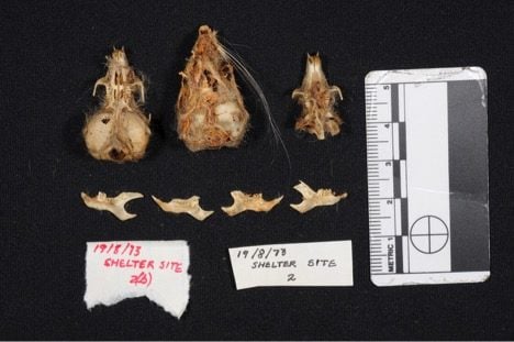
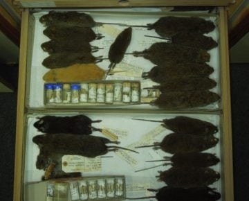
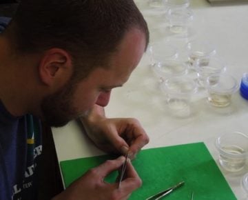
Before data collection can begin, I retrieve the specimens from their respective cabinets. During this process I have come across several interesting specimens (Figure 1). Additionally, as many of the specimens are still articulated with muscular and connective tissue present, some processing is required. For these specimens, a singular hind and forelimb are carefully removed and placed into individual containers filled with water to reconstitute the flesh and make it easier to separate individual skeletal elements (Figure 2). These elements are then carefully cleaned to remove any remaining soft tissue using needle point forceps, dissection micro-scissors, and a lot of patience. Once this is complete, the specimens are placed in an open cabinet to dry.
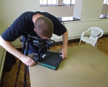
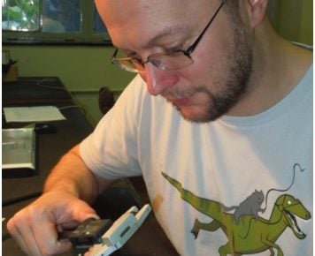
After drying, the specimens are measured using needle point Paleo-Tech Hillson-Fitzgerald dental calipers (Figure 3). This process takes a steady hand, precluding the consumption of the copious amounts of coffee I am accustom to most mornings. Following the linear measurements, the femora and humeri are digitally photographed in standardized positions using a macro lens in order to conduct an outline based analysis (Figure 4).
As doing these same tasks repetitively week after week is not only monotonous, but also can result in sever caffeine withdrawals and thoracic vertebral pain, when a break is needed there are plenty of other things to do. While here I have devoted a portion of my time (yes this is what I like to do for a break) to assessing the microfaunal skeletal part proportions found in various owl pellet accumulations curated at the museum. Again, you never know what interesting things you will find (Figure 5).
With the modern collection almost fully analyzed, next up is the fossils….